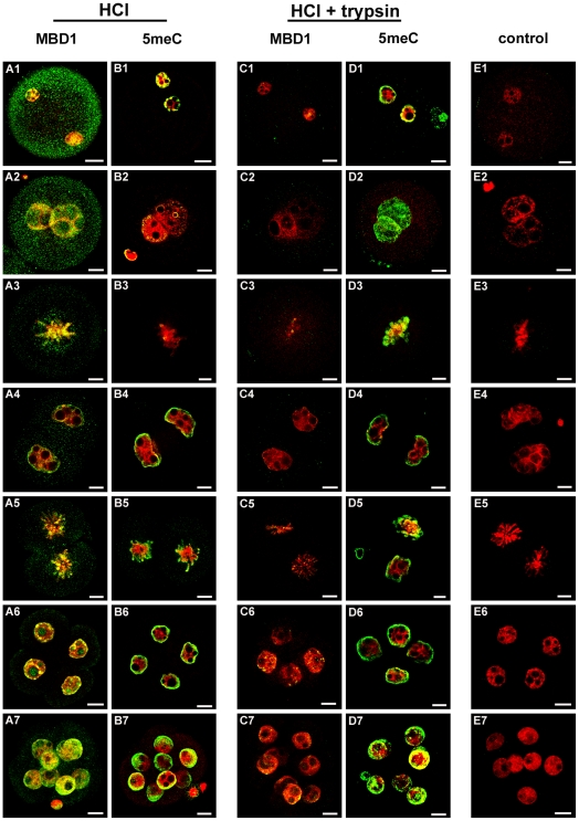
Figure 4. Localization of 5meC in the early embryo by staining for MBD1 and the effect of tryptic digestion on antigenic unmasking of 5meC.
Embryos were freshly collected from the reproductive tract and fixed and antigen unmasked (HCl) as in Fig 1. They were then stained with either anti-MBD1 (MBD1) (A and C ), anti-5meC (5meC) (B and D), non-immune IgG (E) (control). Some embryos were subjected to further antigenic unmasking by tryptic digestion (HCl + trypsin) (C, D, E). Embryos were assessed at the (1) zygotic PN2, (2) PN5, (3) zygotic metaphase, (4) interphase 2-cell, (5) 2-cell metaphase, (6) 4-cell, and (7) 8-cell stages. Each image is a single (0.77 µm) confocal section through embryos except metaphase 2-cell chromosomes and 8-cell embryos which are complied z-stacks of multiple sections. The images shown here are representative for at least seven independent replicate experiments with at least 10 embryos per observation group per replicated. The scale bars are 10 µm.

