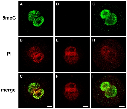
Figure 5. Specificity of 5meC staining.
Zygotes at the PN5 stage were antigenically unmasked by combined acid and trypsin pretreatment and stained with anti-5meC as described in Fig 1, (A-C) or stained with anti-5meC in the presence of excess (0.6 µM ) free 5meC (Sigma) (D-F). To further asses specificity staining with an anti-5meC from an alternative source was performed (G-I). (mouse monoclonal to 5meC, used as 1∶100 dilution and incubated at 4°C for 18 h; Abcam ab73938). This showed the same pattern of staining as was the antibody used in the rest of the study. Representative of three independent replicates.

