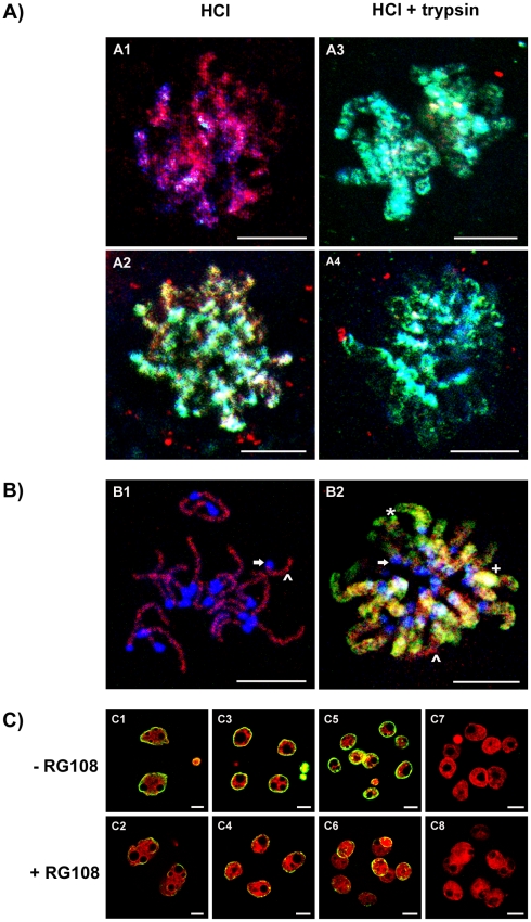
Figure 6. Relative distribution of MBD1 and 5meC staining on metaphase chromosomes.
A) Triple staining of MBD1, 5meC and DNA in 1-cell and 2-cell metaphase chromosomes after antigenic unmasking by acid or acid plus trypsin. Zygotes (A1 and A3) and 2-cell (A2 and A4) embryos were collected at 31 and 53h after hCG, respectively, as they were entering metaphase. They were fixed and treated with acid alone (A1 and A2) or acid followed by acid plus trypsin (HCl + trypsin) (A3 and A4). DNA was counter-stained with DAPI (purple), anti-5meC (FITC, green) and MBD1 (red). Images shown are the merged result of these three channels. Regions of DNA in which anti-5meC and anti-MBD1 are co-localised appear white-yellow, regions of anti-MBD1 alone are pink, and those with anti-5meC alone stain blue-green. Images are z-stacks of multiple confocal sections through the chromosomes. Representative of three independent replicates. The scale bars are 10 µm. B) High resolution image of triple-stained metaphase chromosomes from zygotes and 2-cell embryos. Condensed chromosomes from zygotes (B1) were fixed by the air-dried method [16] and 2-cell condensed chromosomes (B2) by formaldehyde. Images were captured using a 100× oil objective and multiple confocal sections through the chromosomes were collected and the Z-stack images compiled into a two-dimensional representation. Regions of the genome are illustrated as follows: ↑, undecorated DNA; ∧, DNA stained only with anti-MBD1; *, DNA stained only with anti-5MeC; +, DNA dual stained with anti MBD1 and anti-5meC. Representative of three independent replicates. The scale bars are 10 um. C)The role of DNA methyltransferase in the maintenance of 5meC over the first cell cycles. Zygotes (25h post-hCG) were cultured in standard media [35] (- RG108) or media supplemented with RG108 (5 µM) for 16 h. This incubation period covered the time of DNA synthesis in the 2-cell embryo [37]. The embryos were then fixed and stained with anti-5meC (control – C1, RG108 – C2) or the embryos were extensively washed and then cultured for another 24 h (control – C3, RG108 – C4) or 32 h (control – C5, RG108 – C6) prior to staining with anti-5meC. Non-immune controls (control – C7, RG108 – C8). Representative of five independent replicates with at least 10 embryos per treatment dose. The scale bars are 10 µm.

