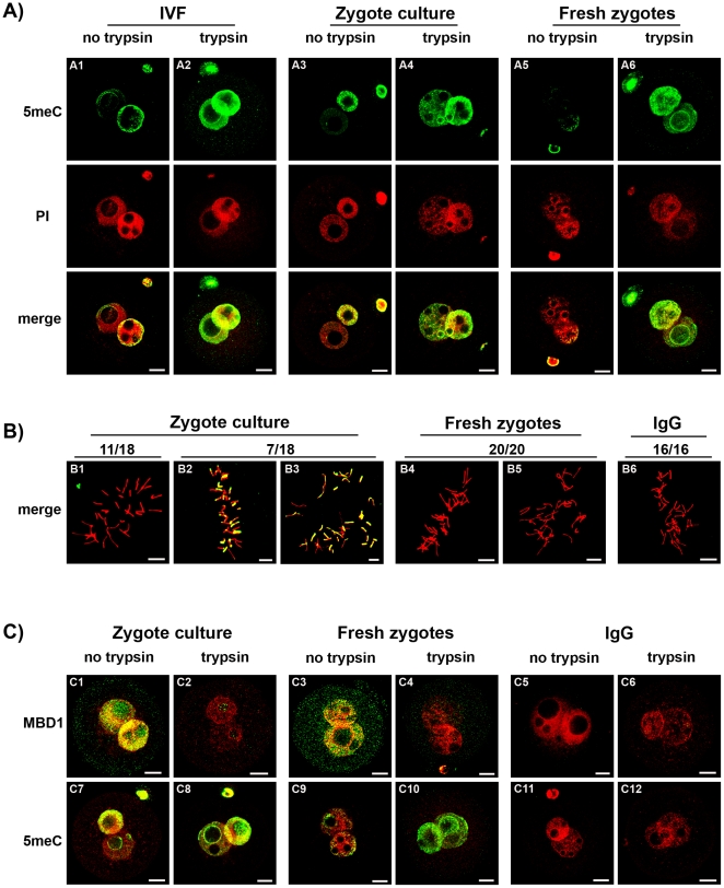
Figure 7. The effect of embryo manipulation on 5meC staining in zygotes.
A)Zygotes were created by routine mouse IVF [40] and culture in vitro (8 h) to the PN5 stage (IVF) (A1 and A2) ; collected after fertilization in the reproductive tract 17 h after hCG and then culture in vitro for 8 h to the PN5 stage (Zygote culture) (A3 and A4), or they were collected directly from the reproductive tract at the PN5 stage, and fixed without further treatment (Fresh zygotes) (A5 and A6). The zygotes were fixed, subjected to acid unmasking and then either buffer (no trypsin) (A1, A3 and A5) or tryptic digestion (trypsin) (A2, A4 and A6) as in Fig 2. Zygotes were stained with anti-5meC (green) or PI (red) and both channels merged. Representative of three independent replicates. The scale bars are 10 µm. Zygotes were prepared as in (A) but were fixed with methanol by the air-dried method [16] and stained during chromosome condensation. In cultured zygotes a proportion showed variable segments of chromosome that stained for 5meC (B1-3). In Fresh zygotes no 5meC staining was observed (B4-5). The proportion of the total number of metaphase zygotes from each treatment that showed the illustrated pattern of staining is shown above the figures. No 5meC staining was observed in no-immune control (B6). The scale bars are 10 µm.

