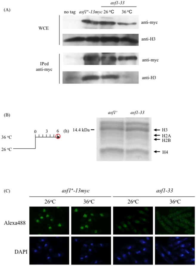
Figure 5. Interaction between Asf1 and histone H3 was lost and Asf1-33 proteins were mislocalized at 36°C in the asf1-33 mutant.
(A) Immunoprecipitation assay to investigate the interaction between Asf1 and histone H3. L972 (asf1+), SKP561-15 (asf1−-13myc-kanr) and SKP605-33 (asf1-33-13myc-kanr) strains were incubated at 26°C and 36°C for 6 h. The cells were collected by centrifugation and washed once with STOP buffer. Two milligrams of total proteins were used. After incubation with Protein G sepharose at 4°C for 1 h, the samples were washed five times with HB buffer. The samples were suspended in SDS-sample buffer and boiled at 100°C. Proteins were detected by western blotting as described in Materials and Methods. (B) Extraction of histone proteins from asf1 mutants. L972 (asf1+) and SKP593-33P (asf1-33-13myc-kan r) strains were incubated at 26°C for 24 h. The temperature was increased to 36°C and cells were incubated for a further 6 h. Extraction of histone proteins was performed as described in the Materials and Methods. Extracted histone proteins were separated by SDS-PAGE and stained with Coomassie Blue. (C) Immunofluorescence images showing the localization of Asf1-13myc in the asf1+ strain and asf1-33 mutant. SKP561-15 (asf1+-13myc-kanr) and SKP605-33 (asf1-33-13myc-kanr) were incubated at 26°C or 36°C for 6 h and immunofluorescence analysis was performed as described in Materials and Methods.

