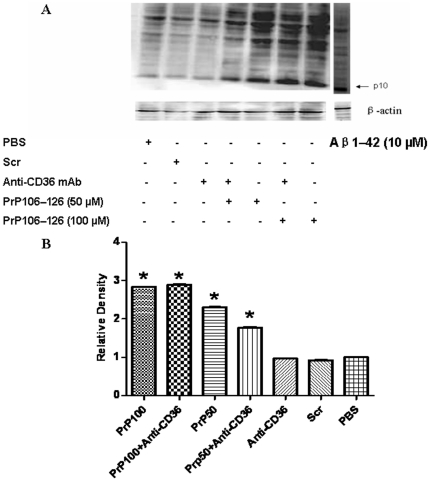
Figure 6. Western blot analysis of caspase-1 cleavage.
BV2 microglia were treated or not with two different concentrations of neurotoxic prion peptides (50 µM and 100 µM, respectively) for 24 h. The role of CD36 in caspase1 cleavage was examined by 1 h-preincubation with anti-CD36 antibody. A. Extracts were prepared and immunoblotted with caspase-1 antibody as described in Materials and Methods. The blot was stripped and reprobed with anti-β-actin antibody to estimate the total amount of protein loaded in gel. Representative blots of caspase-1 and β-actin are shown. Extracts of Aβ1–42-treated cells were used as positive control. B. Bars represent the relative levels of cleaved caspase-1, compared with β-actin, and were expressed as arbitrary units. Data are the means±S.D of three independent experiments. * P<0.05, significantly different from control cells.

