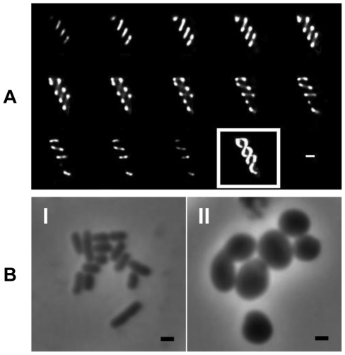
Figure 1. Localization and morphological role of the S. Typhimurium Mre proteins.
(A) Fluorescence microscopy montage showing z-sections taken of MreB-GFP fusions in WT SL1344 revealing a helical distribution. Slices taken at 0.1 µm intervals on live cells in mid log phase going from left to right followed by maximum intensity projection (boxed). (B) Morphology of WT S. Typhimurium (I) and ΔmreC (II) reveal the mutant has changed from rod to round-shaped, with some heterogeneity in size noted. In all images the bar represents 1 µm.

