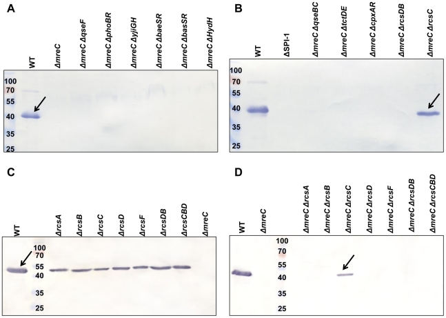
Figure 5. Western blotting screen of ΔmreC two-component system double mutants for recovery of SPI-1 T3S.
Panels A and B show western blots of total protein samples obtained from SL1344 WT, ΔSPI-1, ΔmreC, ΔmreC ΔqseF, ΔmreC ΔphoBR, ΔmreC ΔyjiGH, ΔmreC ΔbaeSR, ΔmreC ΔbasSR, ΔmreC ΔhydH, ΔmreC ΔqseBC, ΔmreC ΔtctDE, ΔmreC ΔcpxAR, ΔmreC ΔrcsDB, and ΔmreC ΔrcsC strains with αSipC antibody. Panels C and D show western blot of total protein samples obtained from SL1344 WT, ΔrcsA, ΔrcsB, ΔrcsC, ΔrcsD, ΔrcsF, ΔrcsDB, ΔrcsCBD and ΔmreC strains along with the ΔmreC ΔrcsA, ΔmreC ΔrcsB, ΔmreC ΔrcsC, ΔmreC ΔrcsD, ΔmreC ΔrcsF, ΔmreC ΔrcsDB, and ΔmreC ΔrcsCBD double mutants with αSipC antibody. SipC is indicated at approximately 43kDa.

