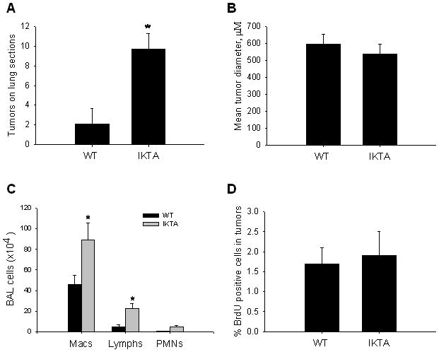
Figure 3. Characterization of lung tumors in WT and IKTA mice.

A) Lung sections from urethane-treated IKTA mice and WT controls treated with doxycycline throughout the course of tumor formation (week -2–16) were sectioned at four predetermined depths and tumors were counted on each H&E stained section. B) Morphometric analysis of tumor diameter was assessed as the mean of 4 separate diameter measurements of all tumors (taken at 450 angles). C) Total number of macrophages (macs), lymphocytes (lymphs), and polymorphonuclear leukocytes (PMNs) in BAL from IKTA and WT mice (n=6 per group, *=p<0.05 compared to WT). D) Bromodeoxyuridine (BrdU) was injected IP 2 hours prior to sacrifice and the percentage of BrdU positive cells per total number of nuclei in tumors was assessed.
