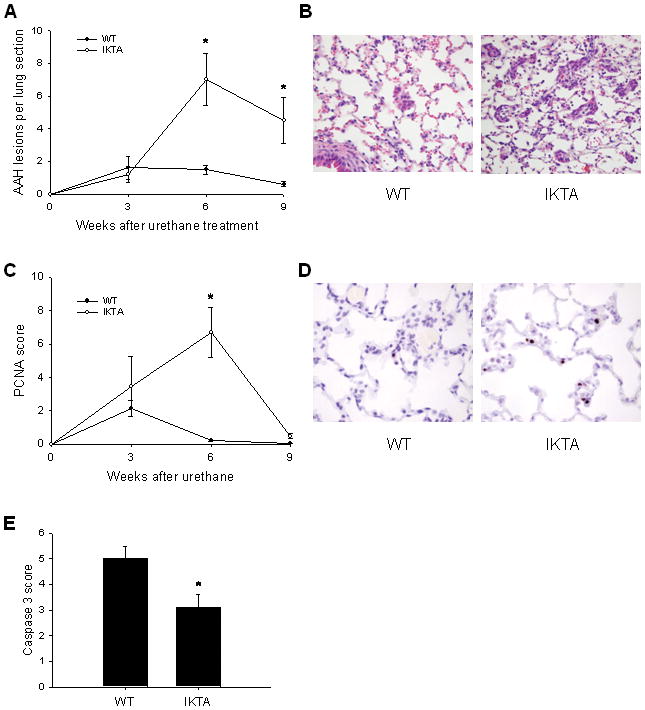
Figure 5. Increased number of atypical adenomatous hyperplasia (AAH) lesions in lungs of IKTA mice is associated with increased proliferation of cells in lung parenchyma.

A) WT and IKTA mice were treated with doxycycline (dox) beginning 2 weeks before urethane and continuing until time of harvest at 3, 6, or 9 weeks post-urethane. AAH lesions were counted on H&E stained lung sections (n=6–12 per group, *=p<0.05 compared to WT mice at the same time point). B) Illustration of characteristic AAH lesions in WT and IKTA mice. C) PCNA scoring was done on H&E stained lung sections. (n=6–12 per group, *=p<0.05 compared to WT at the same time point). D) Immunohistochemistry for PCNA showing nuclear staining (brown) predominantly in type II alveolar epithelial cells throughout lung parenchyma (original magnification x400). E) Active caspase-3 scoring on lung sections from dox-treated WT and IKTA mice treated continuously with dox and harvested at 9 weeks post-urethane (n=8 for IKTA and 10 for WT, *=p<0.05). Semi-quantitative scoring of PCNA or active caspase-3 staining was performed on ten sequential, non-overlapping tissue fields of lung parenchyma evaluated under x400 magnification. Each tissue field was scored using a 0 to 4 point system (0 – no positive cells; 1 – ≤5% positive cells; 2 – 5–10% positive cells; 3 – 10–25% positive cells; 4 – >25% positive cells).
