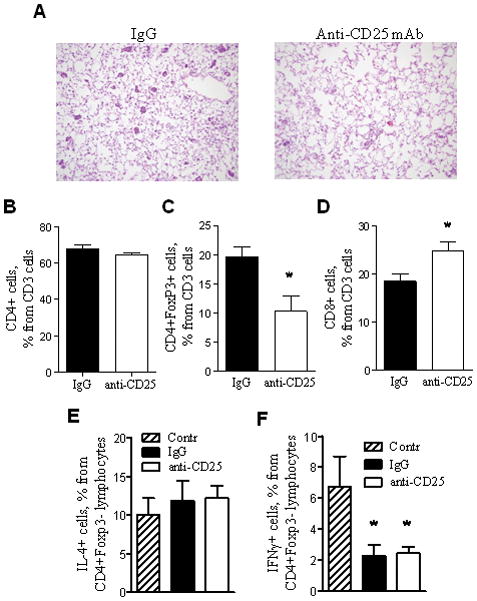
Figure 8. Depletion of Tregs in IKTA mice increases infiltration of lungs with CD8 lymphocytes and reduces the number of atypical adenomatous hyperplasia (AAH) lesions.

A) Photomicrographs of lung sections demonstrating AAH lesions at 6 weeks post-urethane in IKTA mice treated with anti-CD25 antibodies (CD25) or isotype control antibodies (IgG) per protocol (original magnification x200). B–D) Percentages of total CD4+ T cells, CD8+ T cells, and CD4+ Foxp3+ Tregs identified by flow cytometry in lungs from anti-CD25 or isotype control antibody-treated IKTA mice at 6 weeks after injection of urethane (1 week after last antibody dose) (n=4 per group, *=p<0.05). E,F) Percentages of CD4+Foxp3-IL-4+ and CD4+Foxp3-IFNγ+ lymphocytes isolated from lungs of doxycycline-treated IKTA mice at 1 week after a single IP dose of 500 μg of anti-CD25 (PC61) or isotype control antibodies. Cells were re-stimulated with PMA/ionomycin for 6 hours in vitro and analyzed by flow cytometry.
