
Fig. 5.
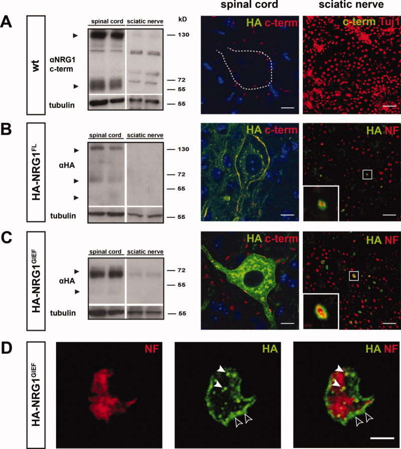
Limited axonal transport of NRG1 type III. (A) (left panel) Endogenous full-length NRG1 (140 kD) and a C-terminal processing product (∼ 60 kD; arrowheads) are detected with an antibody against the C-terminus of NRG1 (αNRG1 C-term) in wt spinal cord protein lysates at P12, whereas corresponding sciatic nerve expression is virtually absent. (right panel) Endogenous NRG1 accumulates on the plasma membrane of spinal cord motor neurons after immunostaining with the αNRG1 C-term antibody. Nuclei are counterstained with DAPI (blue). Corresponding NRG1 immunoreactivity is absent from the sciatic nerve. Sciatic nerve axons are marked by tubulin (Tuj1; red). (B,C) (left panels) When probed with an anti HA-antibody (αHA), HA-NRG1 type III (140 kD) and two N-terminal processing products (∼ 70 kD and ∼ 45 kD; arrowheads) are present in spinal cord protein lysates from HA-NRG1FL transgenic mice at P12, but barely detectable in sciatic nerve. Similarly, HA-NRG1GIEF (∼ 70 kD) and a smaller protein species (∼ 45 kD; arrowheads) are prominently expressed in spinal cord lysates from HA-NRG1GIEF mice, whereas sciatic nerve expression is low. (right panels) Immunostaining for axons (neurofilament 200, NF; red) and the HA-epitope (green) reveals localization of HA-NRG1FL and HA-NRG1GIEF at the surface of individual sciatic nerve axons (insets). Scale bars, 10 μm. (D) Confocal images of an individual sciatic nerve axon in cross section from an adult HA-NRG1GIEF transgenic mouse. Immunostaining for neurofilament 200 (NF; red) and the HA tag (green) reveals expression of HA-NRG1GIEF in vesicle-like structures (white arrowheads) and on the axonal surface (empty arrowheads). Scale bar, 2.5μm. [Color figure can be viewed in the online issue, which is available at wileyonlinelibrary.com.]