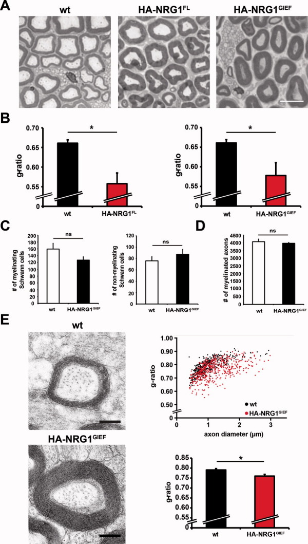
Fig. 6.

HA-NRG1GIEF promotes hypermyelination in the PNS and CNS of transgenic mice. (A) Sciatic nerve hypermyelination in HA-NRG1FL and HA-NRG1GIEF transgenic mice. Methylenblue-Azure II staining for myelin on semithin sciatic nerve cross sections at 2 months of age. Scale bar, 10 μm. (B) Quantification of myelin sheath thickness by g-ratio analysis. HA-NRG1FL and HA-NRG1GIEF transgenic mice exhibit significantly thicker myelin compared with wt (*P < 0.05). Bars represent mean g-ratios ± SEM (n = 3; >100 axons/mouse). (C) The number of myelinated and nonmyelinated Schwann cells is not changed in HA-NRG1GIEF transgenic mice compared with wt at 2 months of age (n = 3; mSchwann cells P = 0.08; nmSchwann cells P = 0.18). Error bars, ±SEM. (D) The total number of myelinated axons in sciatic nerves from HA-NRG1GIEF transgenic mice at 2 months of age is unaltered (n = 3 each; P = 0.29). Error bars, ±SEM. (E) Electron micrographs of the corpus callosum at 2 months of age. Scatter plot (upper panel) displays g-ratios as a function of axon diameter. The average g-ratio (lower panel) in HA-NRG1GIEF transgenic mice is significantly reduced when compared with wt (wt, n = 3; HA-NRG1GIEFn = 4; *P < 0.05). Error bars, ±SEM. Scale bar, 0.25 μm. [Color figure can be viewed in the online issue, which is available at wileyonlinelibrary.com.]
