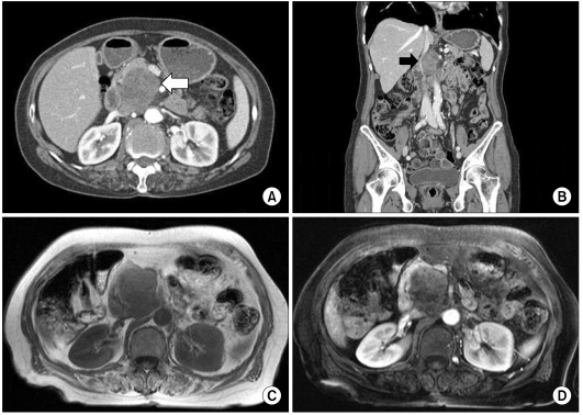
Fig. 1.
Abdominal computed tomography (CT) scan shows growth of the tumor, which is heterogeneously enhanced by contrast media (A, white arrow). CT scan shows the tumor, which is abutting to the inferior vena cava (B, black arrow). Magnetic resonance imaging shows a 5.3 × 4 cm heterogenously enhanced mass (C, D).

