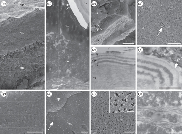
Figure 2.
(a,c,d,g–i) Scanning and (b,e,f,j) transmission electron micrographs of cuticle ultrastructures in fossil beetles with well-preserved metallic colours. (a,b) Vertical sections through the cuticle showing epicuticle (ep), exocuticle (ex) and endocuticle (en). r, resin. (c) Fractured vertical section through the cuticle showing laminated epicuticle (ep) and exocuticle (ex). s, sediment. (d) Detail of surface of lamina in epicuticle showing fibrillar network perforated by pore canals (arrow). (e) Vertical section through the cuticle showing alternating electron-dense and electron-lucent laminae in epicuticle (ep), and uniformly electron-dense laminae in exocuticle (ex). r, resin. (f) Vertical section through the epicuticle showing multi-laminate outer epicuticle (arrow). s, sediment. (g) Vertical section through the exocuticle showing laminations. (h) Oblique fractured section through the epicuticle (ep) and exocuticle (ex) showing arcuate patterns and pore canals (arrow) in the exocuticle. (i) Internal-facing surface of the base of the exocuticle showing pore canals and, inset, pore canal filaments. (j) Membranes of the basal endocuticle (en) and/or basement membrane. Note, granular texture of the endocuticle. Scale bars: (a,b,e) 1 µm; (c,d,g,h) 2 µm; (f,j) 500 nm; (i) 5 µm, inset, 1 µm.

