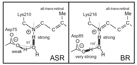
Figure 13.
Schematic drawing of hydrogen bonds of the water molecule locating between the protonated Schiff base and its counterion. A part of all-trans retinal is depicted, β-ionon ring and ethylenic part from C6 to C12 are omitted. The numbers are the angle of the N-O-O atoms derived from the crystal structures of ASR and BR (PDB entries are 1XIO and 1C3W, respectively). Hydrogen bonds are indicated by the dashed lines with their strength. This figure is reprinted with permission from Furutani et al. [20]. Copyright 2005 American Chemical Society.

