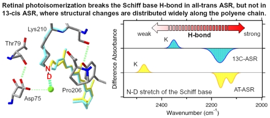
Figure 21.
(Left) The X-ray structure around retinal Schiff base. Yellow retinal is all-trans form and blue retinal is 13-cis form. (Right) The diagram of the ASRK minus ASR infrared spectra in X-D vibration region. It shows only N-D stretch of the Schiff base. This figure is reprinted with permission from TOC of Kawanabe et al [59]. Copyright 2006 American Chemical Society.

