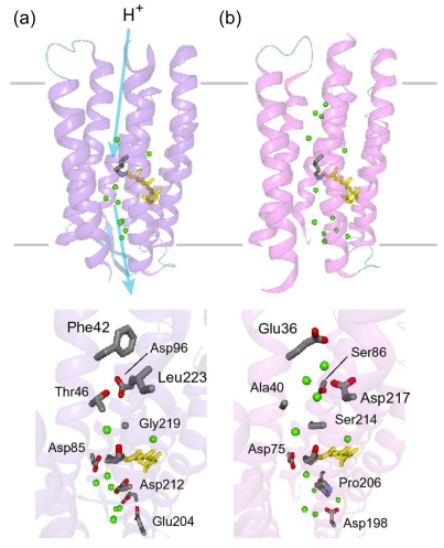
Figure 41.
X-ray crystallographic structures of BR [7] (a) and ASR [16] (b). Top and bottom panels represent views from the membrane plane and the cytoplasmic side, respectively. In the top panel, top and bottom regions correspond to the cytoplasmic and extracellular sides, respectively. The retinal chromophore is colored yellow, and green spheres represent internal water molecules.

