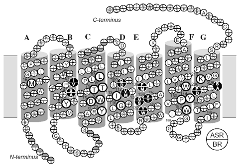
Figure 5.
Comparison of amino acid sequences of ASR and BR. The transmembrane topology is based on the crystallographic three-dimensional structures. The sequence alignment was done using CLUSTAL W [21] with the default settings. Single letters in a circle denote residues common to ASR and BR. The residues that are different in ASR and BR are denoted at the top and bottom of the circles, respectively. The residues forming the retinal binding site within 5 Å of the chromophore are shown by bold or filled circles. This figure is reprinted with permission from Furutani et al. [20]. Copyright 2005 American Chemical Society.

