
Fig. 1.
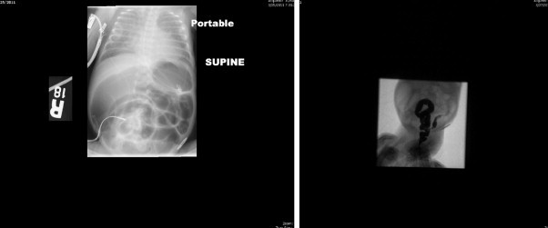
(a) Plain X-ray showing dilated loops of small intestine. Subsequent water soluble contrast enema (b) showed a microcolon that could not be visualized in its entirety.
Official websites use .gov
A
.gov website belongs to an official
government organization in the United States.
Secure .gov websites use HTTPS
A lock (
) or https:// means you've safely
connected to the .gov website. Share sensitive
information only on official, secure websites.
(a) Plain X-ray showing dilated loops of small intestine. Subsequent water soluble contrast enema (b) showed a microcolon that could not be visualized in its entirety.