
Figure 8. In vivo epifluorescence imaging of translocation of BMM loaded with catalase nanozyme across the BBB in a neuroinflamation mouse model.
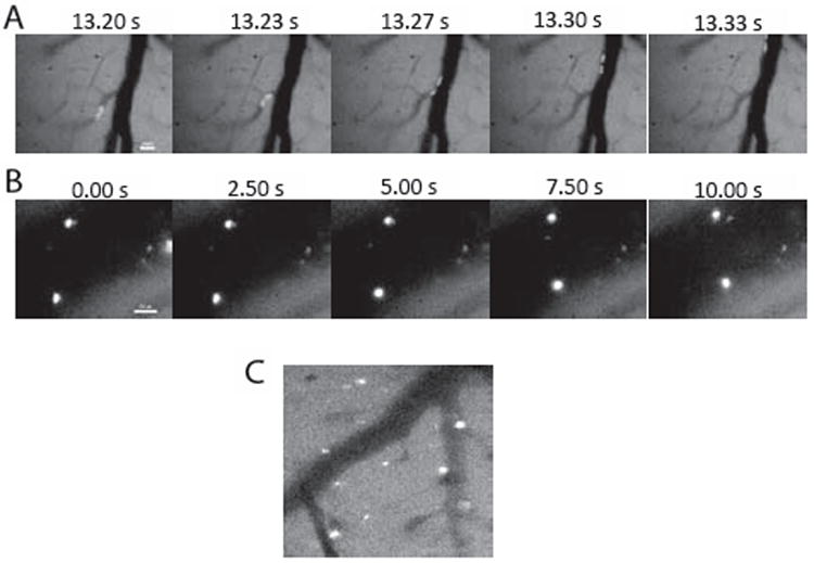
C57BL/6 mice with cranial windows were i.c. injected with 10ug LPS to produce inflammation in the brain area close to the window. Twenty four hours after intoxication, BMM loaded with Alexa 488-labeled nanozyme were adoptively transferred via i.j.v., and images A: immediately after administration; B: 4 hours and C: 24 hours after the injection were taken. Macrophages carrying fluorescently labeled nanozyme first seen moving fast along the microvessels (A), then slow down and adhere to the endothelial wall (B), and finally, were stationary and appeared to have translocated across the BBB into the parenchyma(C). Bar: 20μm.