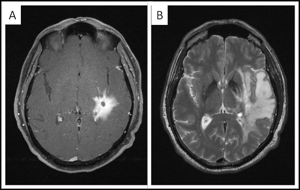
Figure 1.

3 Tesla MRI imaging of the brain. A. Axial fat-saturated postcontrast Tl-weighted image shows the 3.5 × 2.9 cm infiitrative enhancing tumor centered in the left posterior putamen, left posterior subinsular region, and adjacent left mid temporal region. B. Axial T2-weighted image shows extensive associated vasogenic edema, with relatively little mass effect.
