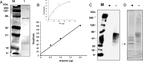
Fig. 4.
(A) SDS–PAGE of the fusion protein galectin-1-hum-β3GN-T2, purified on a UDP-agarose column (1) and marker (M). (B) The catalytic activity of the galectin-1-hum-β3GN-T2 (0.5–2 µg of enzyme) using 50 µM UDP-GlcNAc as the sugar donor substrate. The insert shows the catalytic activity of galectin-hum-β3GN-T2 with UDP-GlcNAc as the donor substrate at different concentrations (10–40 µg), using ASF as an acceptor substrate. Each assay was carried out for 1 h with 2 µg of enzyme. (C) The chemoenzymatic detection of the transferred C2-keto-Glc on ASF after linking the product with AOB, followed by western blotting and chemiluminescence detection. Marker (M) and lanes (+) and (−) represent PNGase F-treated and -untreated samples, respectively. (D) SDS–PAGE analysis of the loading samples of ASF, with and without PNGase F treatment. Arrow points to the PNGase F protein band.

