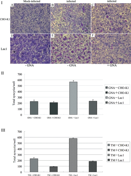
Fig. 6.
NDV-induced syncytium formation in CHO-K1 and Lec1 cells. (A) CHO-K1 and Lec1 cells were pretreated or non-treated with GNA for 1 h at 37°C prior to viral infection, and then infected with NDV (rZJ1-GFP) at an MOI of 1. At 16 hpi, cells were fixed with methanol for 10 min and stained with Giemsa. Syncytia formation was shown in (I). The number of syncytia (cells containing more than three nuclei) was counted in 10 random areas of the well (data are mean ± SD of three independent experiments), as shown in (II). (B) CHO-K1 and Lec1 cells were pretreated or non-treated with tunicamycin (TM) for 24 h at 37°C prior to viral infection, and then infected with NDV (rZJ1-GFP) at an MOI of 1. At 16 hpi, cells were fixed with methanol for 10 min and stained with Giemsa. The number of syncytia (cells containing more than three nuclei) was counted in 10 random areas of the well (data are mean ± SD of three independent experiments), as shown in (III).

