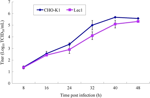
Fig. 8.
Multistep growth curve of NDV infection in CHO-K1 and Lec1 cells. CHO-K1 and Lec1 cells were infected with rZJ1-GFP at an MOI of 0.01, and at the assigned time points post-infection, nearly 300 μL of supernatant was harvested and replenished with the same amount of fresh medium. The virus titers in the collected supernatant were quantitated in triplicate by TCID50 in CEF cells.

