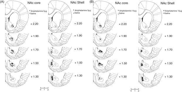
Figure 4.
Histological assessment of cannula placements within the nucleus accumbens. Representative coronal sections from rats that received microinjections of 5.0 µg amphetamine (A) or 10.0 µg amphetamine (B) into the NAc core and NAc shell. Outlines are reproduced from Paxinos and Watson (1998) The Rat Brain in Streretaxic Coordinates, 5th edition with permission from Elsevier)and coordinates refer to the distance in millimetres anterior to bregma.

