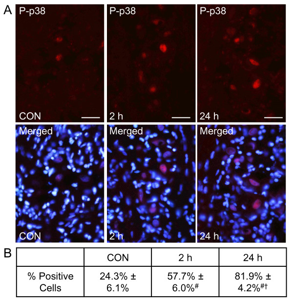
Figure 1.
Increased number of trigeminal ganglia neurons expressing P-p38 in response to CGRP injection into the TMJ capsule. Sections of the posterolateral portion of the ganglion (V3) were obtained from untreated animals (CON), and animals receiving bilateral injections of CGRP after 2 and 24 hours. (A) Images of neuron-satellite glial regions stained for P-p38 are shown in the top panels. Magnification bar = 50 μm. The bottom panels are the same sections co-stained for P-p38 and DAPI. (B) The number of P-p38 positive cells ± SEM for each condition is reported (n = 3 independent experiments) #P < 0.01 when compared to control levels, while † P < 0.01 when compared to CGRP stimulated levels at 2 hours.

