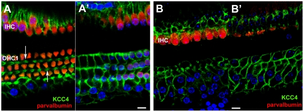
Figure 3. Immunolabelling for KCC4.
A,A'. Normal (undamaged) mature organ of Corti. Confocal images of a whole mount preparation. A, at the level of the cell bodies of the OHC; A', at the level of the cell bodies of the Deiters' cells. Both IHC and OHC are labelled with antibodies to parvalbumin (red). KCC4 labelling (green) delineates the plasma membranes of Deiters' cells, both along the phalangeal processes (arrows in A) and around the cell bodies (A'). KCC4 is also present in the plasma membranes of the supporting cells around the IHC. There is no KCC4 labelling in the region of the pillar cells between the OHC/Deiters' cell region and that of the IHCs. B,B'. 14 weeks post-treatment (CBA/Ca mouse). KCC4 is retained in Deiters' cells in the repaired epithelium. Imaging levels for B and B' approximately the same as for A and A' respectively. Parvalbumin labelling (red) reveals complete absence of OHC but most IHC remain. KCC4 (green) is retained in the plasma membranes of Deiters' cells. The cells' shapes are delineated by the labelling and the variability is apparent. At the level of the phalangeal processes (B) some Deiters' cell are clearly expanded whilst others appear to have retained a thinned phalange. The bodies of many cells are also enlarged and irregular in shape (B') in comparison with the regularity seen in the normal organ of Corti (A'). There is no KCC4 labelling in the pillar cell region. Scale bars: 10 µm.

