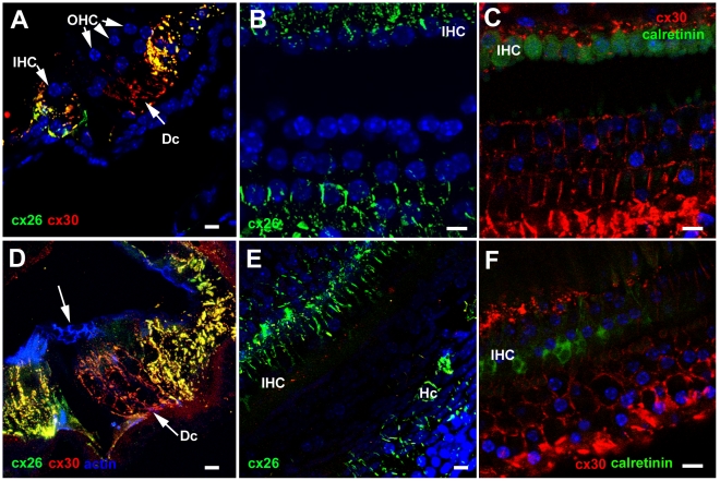
Figure 5. Immunolabelling for Connexin26 and Connexin30.
A–C. Undamaged organ of Corti. A. Frozen section. Cx30 (red) predominates amongst Deiters' cells (Dc), whereas Cx26 labelling (green) is extensively expressed together with Cx30 in cells on the outer (lateral side) of the Deiters' cells (in Hensen's, Claudius and outer sulcus cells) as well as the supporting cells around the IHC and in the inner sulcus. B. Cx26 labelling in whole mount preparation. Large gap junction plaques around Hensen's and Claudius' cells, and inner border and inner sulcus cells are labelled, but little labelling around Deiters' cells. C.Cx30 is present in large gap junction plaques between Deiters' cells. D–F. Repaired organ of Corti following hair cell loss. D. CBA/Ca mouse, 4 days post-treatment. In a frozen section, Cx30 predominates around Deiters' cells, with extensive Cx26 expression, together with Cx30 in cells to the lateral and medial sides of the repaired organ of Corti. Arrow indicates the characteristics pattern of actin labelling in the reticular lamina of the repaired epithelium. E. CBA/Ca mouse, 14 weeks post-treatment. Optical section at approximate level of the cell bodies of Deiters' cells. The pattern of Cx26 labelling is the same as that in the undamaged tissue. F. C57BL/6 mouse 3 weeks post-treatment. Cx30 labelling around the bodies of Deiters' cells. The shapes of the cell bodies, defined by the labelling, are much more irregular than those in the undamaged tissue (panel C) but similar to those defined by labelling for KCC4 and GLAST in the repaired epithelium in Figure 4. Scale bars: 10 µm.

