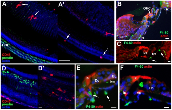
Figure 13. Immunolabelling of Macrophages.
A–C Undamaged organ of Corti. A A'. Immunolabelling for CD45 (red) and for prestin (green) to mark OHC. Focus at the level of the body of the organ of Corti in A and the basilar membrane in A'. In A, macrophages in the nerve tract but none within the organ of Corti itself. Macrophages on the underside of the basilar membrane in A'. Scale bar: 50 µm. B,C. F4-80 in frozen sections. B. Macrophages (indicated by arrows) within the ligament of the lateral wall and in the nerve tract (arrows). C. Macrophages (arrows) on the underside of the basilar membrane. Scale bars: 20 µm. D–F. Following hair cell damage. D D'. C57BL/6 at 24 hours post treatment. D. Focus at the level of the body the organ of Corti shows debris of degenerating OHC labelled for prestin (green). There are no cells labelled with macrophage marker CD45 at this level indicating they are absent from the body of the organ of Corti at the time when hair cell degeneration is occurring. D'. Focus at the level below the organ of Corti reveals CD45 positive cells (red), remain on the underside of the basilar membrane. Scale bar: 20 µm. E. CBA/Ca mouse; 48 hours post treatment. Frozen section labelled for cells expressing F4-80 (green). Macrophages are present on the underside of the basilar membrane and in the nerve tract (arrows) but are absent from the body of the organ of Corti at a time when OHC loss is on-going. Scale bar: 10 µm. F. CBA/Ca mouse 7 days post-treatment. Frozen section labelled for cells expressing F4-80. All OHC have been lost and all debris cleared by this time. Large cell of irregular shape expressing the macrophage marker F4-80 (green) is within the tunnel of Corti. Scale bar: 10 um.

