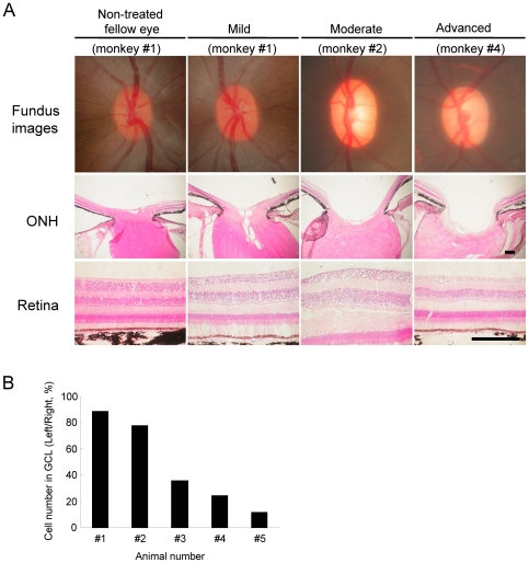
Figure 1. Ocular fundus photographs and histological sections of the retina at each experimental glaucoma stage in laser-treated eyes of cynomolgus monkeys.
(A) Ocular fundus photographs were taken just before the end of the experiment for each monkey Each scale bar indicates 200 µm for ONH and retina. (B) The cell numbers in ganglion cell layers (GCL) of each experimental glaucoma stage were estimated by counting GCL cells at a distance between 1750 and 2200 µm from the optic disc. Cell number was expressed as (healthy cell number on GCL of left eye)/(healthy cell number on GCL of right eye)×100% for each animal.

