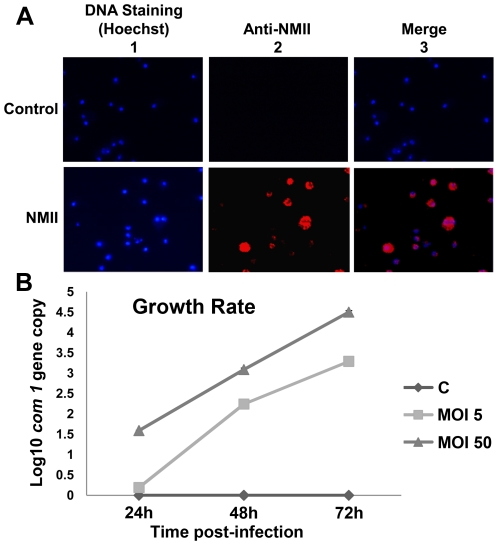
Figure 1. Growth rate and indirect immunofluorescence assay (IFA) staining of NMII in THP-1 cells.
Cells were infected with NMII at MOI of 50 for 24 h. Infected cells were then washed and incubation was continued for 48 h. Infected and uninfected THP-1 cells were fixed with paraformaldehyde and permeabilized. Intracellular C. burnetii were stained by IFA with rabbit anti-Coxiella polyclonal antibodies and viewed using a fluorescence microscope. Panel 1A, IFA staining of NMII infected THP-1 cells. Upper panel: uninfected control cells; lower panel: NMII infected cells. 1 Hoechst staining for host cell DNA; 2 Cells stained with anti-Coxiella antibodies; and 3 Merge. Panel 1B, growth rate of NMII in THP-1 cells. NMII infected and uninfected monocytic THP-1 cells were directly lysed at 24, 48 or 72 h post infection. DNA was extracted and used as a template to quantify the number of C. burnetii com1 gene copies by real-time PCR.

