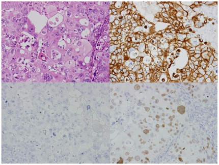
Figure 3. An adenocarcinoma showing typical presentation of the lung profile by immunohistochemistry.
Hematoxylin-eosin stain (left upper), cytokeratin (CK)7 (right upper), CK20 (left lower), and thyroid transcription factor-1 (TTF-1) (right lower). CK7 and TTF-1 are positive in the cytoplasm and nuclei of tumor cells, respectively (original magnification ×200).

