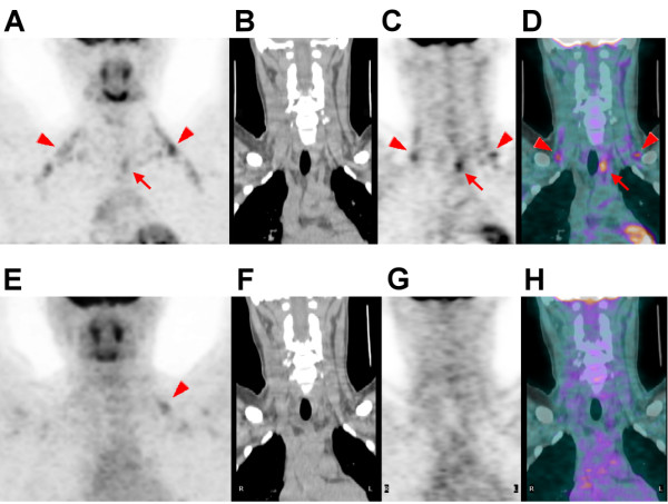
Figure 3.

18F-FDG PET/CT scan (top row) of an 18-year-old man with a history of papillary thyroid cancer treated with a bilateral total thyroidectomy showing activated BAT deposits in the bilateral posterior neck (PN) and supraclavicular (SC) areas (arrowheads), and a hypermetabolic nodule in the left paratracheal area (arrows). The nodule was surgically excised and proved to be a metastatic lymph node. At that time, the TMA of the BAT was 46.6, the neoplastic status score was 3, the outdoor daily average temperature on the date of the PET/CT study was 25.4 °C, the patient's BMI was 29.0 kg/m2, and his fasting blood sugar was 86 mg/dL. On the follow-up 18F-FDG PET/CT scan (bottom row) after the lymph node had been surgically excised, there was no abnormal tumor uptake and only minimal activated BAT deposits in the SC areas (E, arrowhead). At that time, the TMA of the BAT was 9.2, the neoplastic status score was 1, the outdoor daily average temperature on the date of the PET/CT study was 24.4 °C, the patient's BMI was 28.1 kg/m2, and his fasting blood sugar was 99 mg/dL. (A, E: maximum intensity projections; B, F: coronal CT images; C, G: coronal PET images; D, H: coronal PET/CT fusion images).
