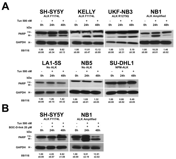
Figure 5.
Activation of the Poly-ADP-ribose-polymerase (PARP) protein. Cells were treated for 48 h with: (A) 500 nM tunicamycin only; (B) 500 nM tunicamycin and 20 μM of the pan-caspase inhibitor BOC-D-fmk. GAPDH was used as sample loading control. Quantification of western blot bands was performed by using ImageJ software [13] as described in Figure 1, except the ratio was calculated between the cleaved and the uncleaved form of PARP.

