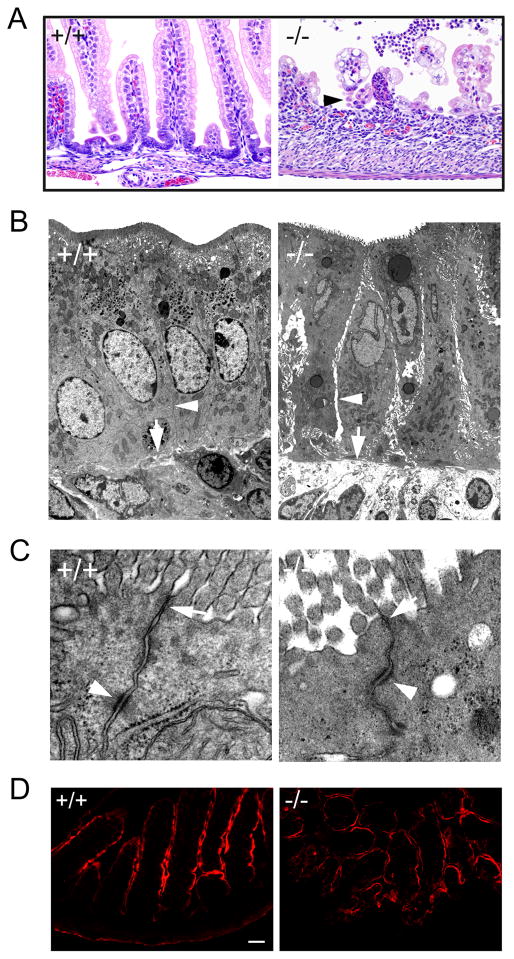
Figure 5.
Disruption of cell-cell adhesion in Cldn7−/− intestines. (A) Light micrographs of H&E stained day P5 Cldn7+/+ (+/+) and Cldn7−/− (−/−) small intestines. The arrowhead in (−/−) pointed to the sloughing epithelial cells. Magnification: ×400. (B) Electron micrographs of day P5 Cldn7+/+ (+/+) and Cldn7−/− (−/−) small intestines. The arrowhead in (−/−) pointed to the intercellular gap along the Cldn7−/− lateral membrane compared to that of Cldn7+/+ (+/+, arrowhead). The arrow in (−/−) revealed the loosening of cell-matrix connection in Cldn7−/− versus the close contact between cell and matrix in Cldn7+/+ intestines (+/+, arrow). (C) The arrows in (+/+) and (−/−) pointed to the apical TJ. The arrowheads indicated the desmosome. Magnifications: (B), ×5,000; (C), ×50,000. (D) Frozen sections of Cldn7+/+ (+/+) and Cldn7−/− (−/−) small intestines were immunostained with an apical protein marker, Ezrin. Bar: 40 μm.

