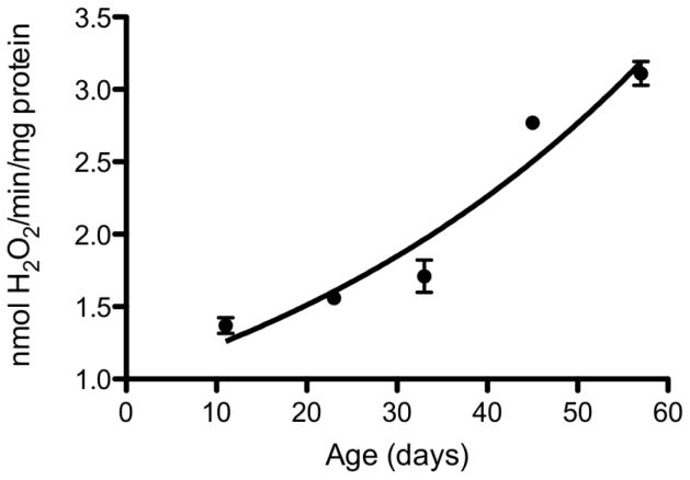
Fig. 2.
Rates of H2O2 release by mitochondria of Drosophila melanogaster at different ages. Rate of H2O2 release was measured in isolated flight muscle mitochondria as an increase in fluorescence due to oxidation of p-hydroxyphenylacetate and the coupled reduction of H2O2 by horseradish peroxidase, using alpha-glycerophosphate as a substrate. From 233.

