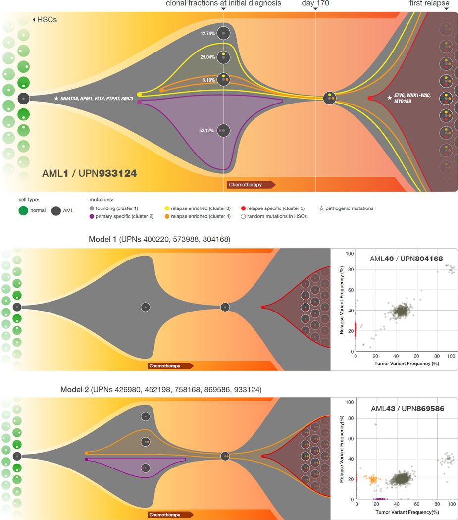
Figure 2. Graphical representation of clonal evolution from the primary tumor to relapse in UPN 933124, and patterns of tumor evolution observed in 8 primary tumor and relapse pairs.
a) The founding clone in the primary tumor in UPN 933124 contained somatic mutations in DNMT3A, NPM1, PTPRT, SMC3, and FLT3 that are all recurrent in AML and probably relevant for pathogenesis; one subclone within the founding clone evolved to become the dominant clone at relapse by acquiring additional mutations, including recurrent mutations in ETV6 and MYO18B, and a WNK1-WAC fusion gene. HSC: hematopoietic stem cell. b) Examples of the two major patterns of tumor evolution in AML: Model 1 shows the dominant clone in the primary tumor evolving into the relapse clone by gaining relapse-specific mutations; this pattern was identified in 3 primary tumor and relapse pairs (UPNs 804168, 573988, and 400220). Model 2 shows a minor clone carrying the vast majority of the primary tumor mutations survived and expanded at relapse. This pattern was observed in 5 primary tumor and relapse pairs (UPNs 933124, 452198, 758168, 426980, and 869586).

