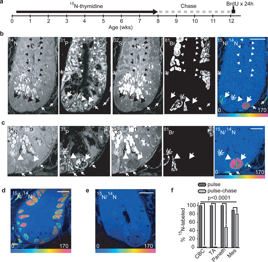
Figure 2. No label-retaining stem cells in the small intestinal crypt.
- 15N-thymidine administered from post-natal day 4 - week 8. After 4-wk chase, BrdU was administered (500 µg i.p. Every 6 h) for 24 hrs before sacrifice (See Supplemental Fig 8).
- 14N: crypt structure and intense signal in intracytoplasmic Paneth granules at the crypt base. 31P: intense intranuclear signal. 32S: intense signal within cytoplasmic Paneth cell granules. 81Br: direct measure of BrdU incorporation. 15N/14N HSI: 15N-thymidine labeling within 15N+/BrdU- Paneth cells (large arrow) and mesenchymal cells (small arrows). No other cells reveal 15N retention. Large arrow head: recently formed (15N−/BrdU+) Paneth cell. Small hatched arrow (middle left side of the crypt): unlabeled Paneth cell, (15N−/Br-). Scale bar=15µm.
- Continued analysis of the same crypt after narrowing the acquisition field. High 15N-signal in a BrdU− Paneth cell (large arrow) and mesenchymal cells closely associated with the crypt (small arrows). BrdU+ CBC (hatched arrow) and Paneth cell (arrow head) are 15N−. Scale bar=5µm.
- 15N/14N HSI image of small intestinal crypt at the end of 15N-thymidine pulse. All nuclei are labeled. Nuclei with lesser degrees of labeling likely represent cells born during a period of lower circulating 15N-thymidine as expected given the different labeling protocols (Supplemental Fig 8). Scale bar=20µm.
- Unlabeled mouse image. The entire crypt contains the natural abundance of 15N. Scale bar=10µm.
- Mean % 15N+ cells at the completion of pulse and after pulse-chase (± standard error of CBC, TA, Paneth, and mesenchymal (Mes) cells (n=3 mice per group).

