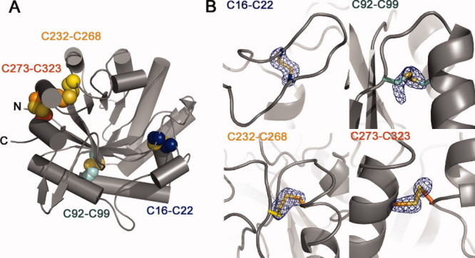
Figure 2.

Disulfide bonding patterns in Hj_Cel5A. (A) Cartoon representation of the protein highlighting positions of the four intramolecular disulfide bonds detected in the electron density. (B) Fo-Fc cysteine sidechain omit maps contoured to 5 σ. Sidechain atoms from the Cβ to the end of the sidechain were deleted from the model before map generation.
