Figure 1.
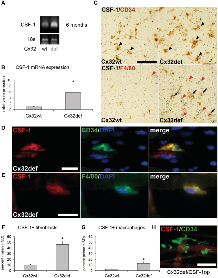
Increased expression of CSF-1 in peripheral nerves of Cx32def mice. (A and B) Semi-quanitiative real-time polymerase chain reaction for CSF-1 messenger RNA and 18S ribosomal RNA expression as endogenous control revealed increased CSF-1 levels in sciatic nerves from 6-month-old Cx32def mice compared with age-matched wild-type (wt) mice (n = 4). Student's t-test *P < 0.05. (C) CSF-1 specific in situ hybridization in combination with immunohistochemistry against CD34 after 10 weeks (top) or F4/80 after 20 weeks (bottom) of exposure on cross-sections of femoral quadriceps nerves from 6-month-old wild-type or Cx32def mice. Arrowheads demarcate accumulations of silver grains in CD34-positive cell profiles and arrows indicate accumulations in F4/80-positive cell profiles. Note accumulation of grains on F4/80-negative ‘ghost profiles’, most probably representing endoneurial fibroblasts (red arrowheads) since these were the only cells showing accumulations after 10 weeks. Scale bar = 50 µm. (D) Double immunocytochemistry against CSF-1 (red) and CD34 (green) or (E) F4/80 (green) on teased nerve preparations (sciatic nerve) from 6-month-old Cx32def mice. Scale bars = 20 µm. (F) Quantification of CSF-1-positive (CD34-positive) fibroblasts or (G) CSF-1-positive (F4/80-positive) macrophages in teased nerve preparations (quadriceps nerve) from 6-month-old wild-type and Cx32def mice (n = 3). Note that fibroblasts are the major CSF-1-expressing cell type. Mann–Whitney U-test *P < 0.05. (H) Double immunocytochemistry against CSF-1 (red) and CD34 (green) on teased nerve preparations (sciatic nerve) from 6-month-old Cx32def/Csf1op mice demonstrated absence of CSF-1 immunoreactivity. Scale bar = 30 µm.

