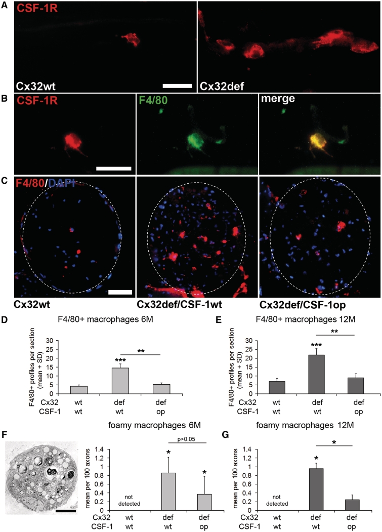
Figure 2.
CSF-1 mediates increase of macrophage numbers in peripheral nerves of Cx32def mice. (A) Immunocytochemistry against CSF-1R (c-Fms; red) on teased nerve preparations (sciatic nerve) from 6-month-old wild-type and Cx32def mice. Scale bar = 30 µm. (B) Double immunocytochemistry against CSF-1R (red) and F4/80 (green) on teased nerve preparations (sciatic nerve) from 6-month-old Cx32def mice. All CSF-1R-positive cell profiles were F4/80-positive macrophages. Scale bar = 30 µm. (C) Immunohistochemistry against F4/80-positive macrophages in cross-sections of femoral quadriceps nerves from 6-month-old wild-type, Cx32def/Csf1wt and Cx32def/Csf1op mice. Broken lines demarcate the boundaries of quadriceps nerves. Scale bar = 50 µm. (D) Quantification of F4/80-positive macrophages in 6-month-old (n = 4–5) and (E) 12-month-old (n = 3–4) Cx32/Csf1 mutants revealed attenuated elevation of macrophage numbers in Cx32def/Csf1op compared with Cx32def/Csf1wt quadriceps nerves. Student's t-test **P < 0.01; ***P < 0.001. (F) Electron microscopy and quantification of foamy macrophages in quadriceps nerves from 6-month-old (n = 4) and (G) quantification of foamy macrophages in quadriceps nerves from 12-month-old (n = 3–4) Cx32/Csf1 mutants. Scale bar = 2 µm. Mann–Whitney U-test *P < 0.05.

