Figure 6.
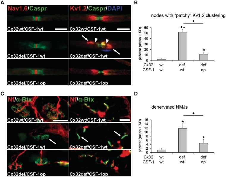
CSF-1 deficiency leads to reduced formation of maldistributed juxtaparanodal ion channels and reduced denervation of neuromuscular junctions in Cx32def mice. (A) Double immunocytochemistry against voltage-gated Na+ channels (Nav1.6; red) or voltage-gated K+ channels (Kv1.2; red) in combination with Contactin-associated protein (Caspr; green) on teased fibre preparations of quadriceps nerves from 6-month-old wild-type, Cx32def/Csf1wt and Cx32def/Csf1op mice. Nodal (Nav1.6) and paranodal (Caspr) domains appeared normally organized in mice of all genotypes, while juxtaparanodal K+ channels frequently presented with ‘patchy’ maldistribution in nerves from Cx32def/Csf1wt mice (arrows). This abnormal distribution of K+ channels causes ‘gaps’ in the immunopositive aspects (arrowheads) and is substantially reduced in the absence of CSF-1. Note the nucleus of an unidentified cell (a putative macrophage) in proximity to the abnormal juxtaparanodal domain. Scale bars = 20 µm. (B) Quantification of nodes of Ranvier with ‘patchy’ K+ channel distribution in quadriceps nerves from 6-month-old wild-type, Cx32def/Csf1wt and Cx32def/Csf1op mice (n = 3–5) revealed reduced formation of ‘patchy’ clustering in Cx32def mice in the absence of CSF-1. Mann–Whitney U-test *P < 0.05; **P < 0.01. (C) Visualization of neurofilament-positive (red) presynaptic axon terminals and α-bungarotoxin-positive (green) postsynaptic terminals in whole mount preparations (left) or cross-sections (right) of flexor digitorum brevis muscles from 12-month-old wild-type, Cx32def/Csf1wt and Cx32def/Csf1op mice. Arrows demarcate neurofilament-negative, denervated (or partially denervated) postsynapses. Scale bars = 30 µm. (D) Quantification of denervated neuromuscular junctions (NMJs) in cross-sections of flexor digitorum brevis muscles from 12-month-old wild-type, Cx32def/Csf1wt and Cx32def/Csf1op mice (n = 3–4) revealed reduced denervation in Cx32def mice in the absence of CSF-1. Mann–Whitney U-test, *P < 0.05.

