Figure 7.
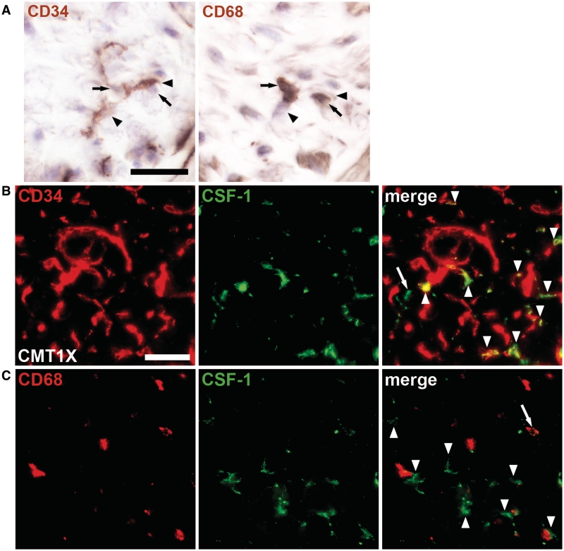
CSF-1-producing fibroblasts are in close contact with macrophages in peripheral nerves of human patients with Charcot–Marie–Tooth type 1. (A) Immunohistochemistry against CD34-positive endoneurial fibroblasts (left; arrowheads) and CD68-positive macrophages (right; arrows) in adjacent serial sections of a frozen sural nerve biopsy from a 47-year-old patient with Charcot–Marie–Tooth type 1X (Table 1, Patient X3) counterstained with haematoxylin. Fibroblasts were in close contact with cells that were identified as macrophages in the adjacent section and vice versa. Scale bar = 20 µm. (B) Double immunohistochemistry against CD34 (red) and CSF-1 (green) on sections of the same biopsy as shown in A. The majority of CSF-1-positive profiles were CD34-positive fibroblasts (arrowheads) while only few profiles, putative macrophages, did not colocalize with CD34 (arrow). Scale bar = 30 µm. (C) Double immunohistochemistry against CD68 (red) and CSF-1 (green) confirmed that only few macrophages were CSF-1-positive (arrow) while the majority of CSF-1-positive profiles, putative fibroblasts (arrowheads), were not CD68-positive but located in close proximity to macrophages.

