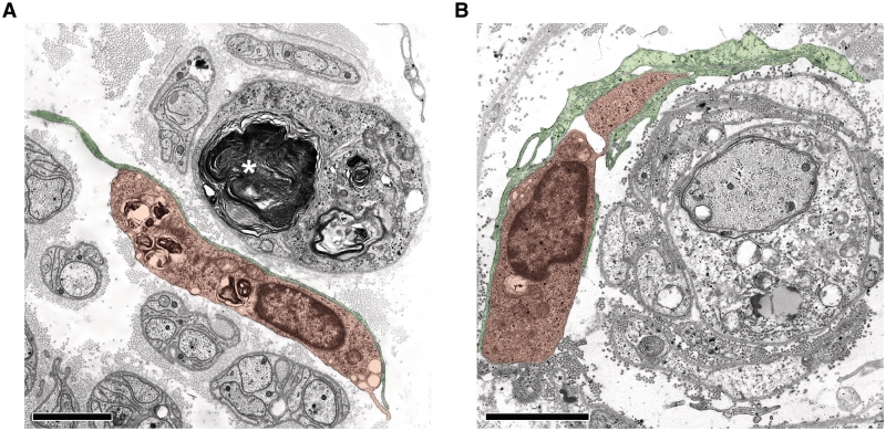
Figure 8.
Endoneurial fibroblasts form direct cell–cell contacts with macrophages in human patients with Charcot–Marie–Tooth type 1. (A) Electron microscopy of a direct cell–cell contact between a fibroblast process (green) and a macrophage containing myelin debris (orange) in a sural nerve biopsy from a patient with Charcot–Marie–Tooth type 1X (Patient X2, Table 1). Note myelin debris in an endoneurial tube devoid of an axon (asterisk). Scale bar = 1.5 µm. (B) Electron microscopy of direct cell–cell contacts between fibroblast processes (green) and a macrophage (orange) in proximity to a demyelinated axon in a sural nerve biopsy from a patient with Charcot–Marie–Tooth type 1A (Patient A1, Table 1). Note that the associated macrophage-fibroblast unit is surrounding the demyelinated axon and Schwann cells profiles reminiscent of onion bulbs. Scale bar = 1.5 µm.

