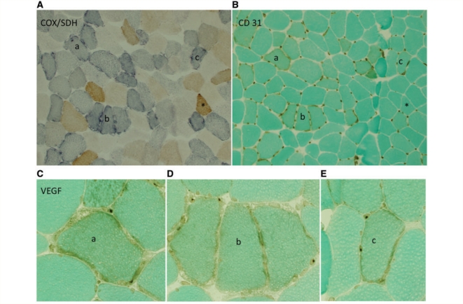
Figure 3.
Capillaries are most abundant around respiration-deficient fibres within the muscle of patients with mitochondrial myopathy. (A) Serial sections within Patient 5 illustrate cytochrome oxidase-positive (brown) and respiration-deficient, cytochrome oxidase-negative, succinic dehydrogenase-positive (intense blue) fibres. (B) Serial section with CD31 immunostaining reveals increased capillaries around cytochrome oxidase-negative compared with cytochrome oxidase-positive fibres. (C–E) VEGF immunostaining shows more intense staining of endothelial perinuclei surrounding individual cytochrome oxidase-deficient fibres. Individual cytochrome oxidase-negative fibres in the serial section are marked as a, b and c; a single cytochrome oxidase-positive fibre is marked with an asterisk. Images in A and B = ×10 magnification, C–E = ×40 magnification. COX = cytochrome oxidase; SDH = succinic dehydrogenase.

