
Figure 1.
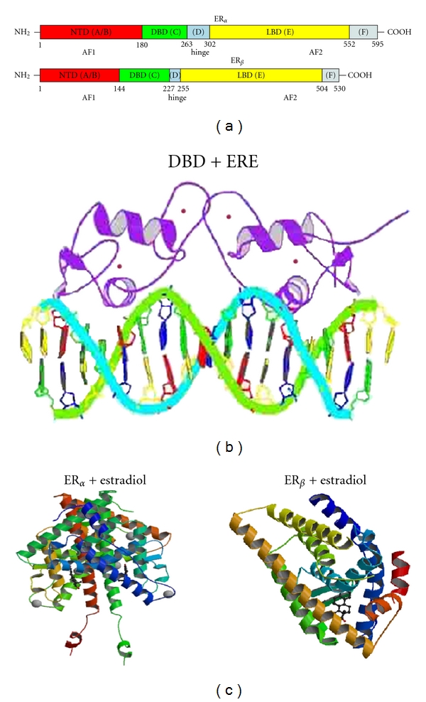
(a) shows the sequence organization of the two isoforms of estrogen receptors, ERα and ERβ. Different domains are highlighted in different colors: NTD—amino terminal domain—in red; DBD—DNA binding domain—in green; hinge region—in blue; LBD—ligand-binding domain—in yellow; F region located towards the C-terminal end—in grey. Amino acid sequence position is given for each domain. (b) shows estrogen receptor DBD in complex with DNA-ERE (estrogen response element). Structure 1HCQ from PDB (protein databank) [13]. (c) shows 3-dimensional structures of ERα (left) and ERβ (right) bound to estradiol (PDB structures 1A52 [14] and 3OLS [15]).