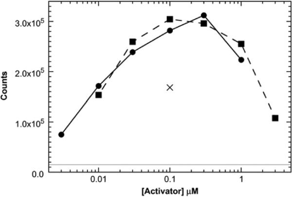
Fig. 1.
Activation of PKR by dp8 (squares; broken line) and dp6 (circles; continuous line). Activation of PKR by 100 nM of a 40-bp dsRNA (×) is included for comparison. The continuous horizontal line represents the background autophosphorylation of PKR in the absence of activator. Activation assays were performed at 100 nM PKR in P50 buffer [20 mM Hepes (pH 7.5), 50 mM KCl, 5 mM MgCl2, and 0.1 mM tris(2-carboxyethyl)phosphine] for 20 min at 32 °C. Reactions contained 0.4 mM ATP and 2 μCi of [γ-32P]ATP. Figure S1 shows the PhosphorImager gel scan used to generate the data in this figure.

