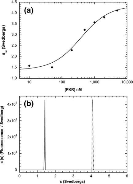
Fig. 3.
bdp8 binding to PKR analyzed by fluorescence-detected sedimentation velocity. (a) PKR titration. Samples contained 15 nM bdp8 and variable concentrations of PKR. Weight-average sedimentation coefficients (sw) were obtained by the integration of g(s*) distributions and were fitted to a hyperbolic binding model to give the following best-fit parameters: Kd=387±89 nM, sw(bdp8)=1.38±0.10 S, and sw(bdp8–PKR complex)=4.35±0.16 S. (b) c(s) distributions of 250 nM bdp8 ( ) and 250 nM bdp8+10 Eq of PKR (----). Conditions: rotor speed, 50,000 rpm; temperature, 20 °C; fluorescence optics.

