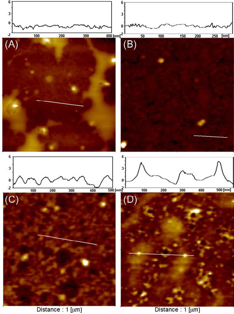
Figure 3.
AFM analysis of neutravidin deposition on glass substrates modified with mono- and bi-functional silanes. (A–B) Surface topography of glass substrates modified with acrylated silane before (A) and after (B) physical adsorption of neutravidin. (C–D) Surface topography of thiol/acrylate silane layer before (C) and after (D) after covalent attachment of neutravidin. Note presence of particles similar in size (6 nm) to neutravidin on glass substrates modified with mixed silane layer.

